Exploring 5 Healthcare Imaging Techniques: Uses and Limitations
Advanced imaging techniques play a critical role across healthcare, with a hand in diagnostics and disease monitoring.
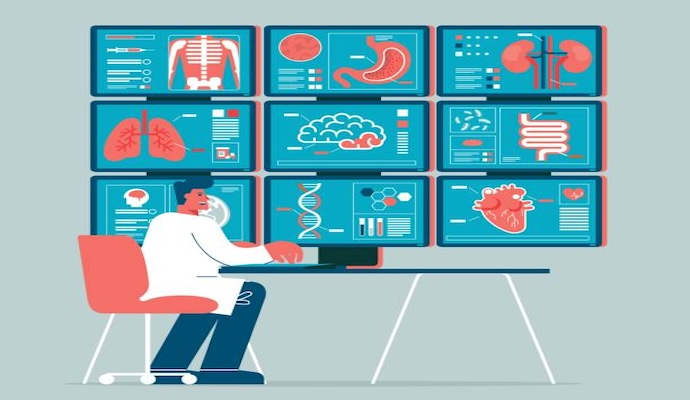
Source: Getty Images
- Advancements in healthcare imaging technologies and techniques have revolutionized diagnostics and disease monitoring. As these tools continue to develop and evolve, healthcare providers can leverage them for early disease diagnosis, periodic patient monitoring, and tracking treatment outcomes.
Despite the benefits and uses of these tools, many have financial or resource-based limitations that hinder their widespread and regular use. Even so, researchers are working to enhance these techniques, improving their accuracy and utility.
In this article, LifeSciencesIntelligence will explore five standard imaging techniques in healthcare and introduce some new developments in medical imaging.
Magnetic Resonance Imaging (MRI)
Magnetic resonance imaging (MRI) is one of the most recognized types of medical imaging. In this technique, the MRI machine — which is cylindrical — emits a magnetic field and computer-generated radio waves to craft detailed images of organs and tissues.
According to Johns Hopkins Medicine, on a molecular level, MRI scans are produced because the magnetic field generated by the machine forces atoms in the body to align in a particular direction; however, the computer-generated waves move the atoms out of these positions. Once the frequencies are turned off, the atoms return to their magnet-oriented positions and emit radio signals, which the computer processes and converts into images of the tissues.
This noninvasive imaging process can provide a look at organs, bones, muscles, and blood vessels. One of the primary benefits of MRIs is that they do not involve any radiation exposure, making it generally safe for patients.
The additional benefits of MRI include soft tissue contrast, multiplanar imaging, and the ability to assess tissue function, blood flow, and more through functional MRI techniques.
Unfortunately, there are some drawbacks to MRI. For example, patients with metal implants, including pacemakers and cochlear implants, cannot be imaged using MRIs because of the strong magnetic fields.
Additional MRI limitations include cost and availability, longer scan times, and limited applications in acute trauma settings. Additionally, these machines require many resources to maintain. For example, MRI machines use helium for cooling properties to ensure that machines remain cold enough to maintain superconductivity; however, in 2022, many helium suppliers started to ration the resource due to limitations. Until an alternative, sustainable cooling method is developed, MRI scans should be used frugally to limit resource usage.
Computed Tomography
Another commonly used medical imaging type is computed tomography (CT) scans, which combine a more straightforward imaging technique, X-rays, with computer processing to image multiple body parts, providing a visual of bones, blood vessels, and organs.
In traditional X-rays, energy beams are targeted at a specific body party and captured on a plate behind the body part for imaging. However, CT scans move X-ray beams around the body to extract similar information and depict an image of the intended body part.
Additionally, newer CT technologies have used computer software to develop 3D images.
Unlike MRIs, CT scans require patients to be exposed to some level of ionizing radiation. Although the exposure is usually limited, it carries a risk of DNA damage, which can result in other issues, including cancers.
Beyond the dangers of ionizing radiation, some patients undergoing CT scans may be given contrast dyes for better visualization. Contrast dyes carry their own risks because tolerability and allergies vary from person to person.
Despite CT scans' limitations, they provide many advantages, including rapid imaging, detailed visualization, multiplanar imaging, and simultaneous assessment.
Positive Emission Tomography
Positive emission tomography (PET) is a nuclear imaging technique where patients are injected with a small radioactive substance, called a radiopharmaceutical product or tracer, that emits positrons as it begins to decay in the body.
Once a positron interacts with an electron, the particles annihilate and emit two gamma rays, also known as photons. These photons travel in opposite directions and are detected by the PET machine and transformed into 3D images using computational technology.
Because PET scans use radioactive substances, there is a small radiation risk. However, radioactive tracers used in PET scans are generally short-lived and decay quickly. They provide sufficient imaging time while minimizing radiation exposure to the patient.
Single-Photon Emission Computed Tomography
Single-photon emission computed tomography (SPECT) is a unique type of imaging that combines strategies used in CT and PET scans. According to the American Heart Association, SPECT scans are non-invasive nuclear imaging tests that use radioactive tracers to show how well organs function.
The machine detects the gamma rays emitted by the radiopharmaceutical product, and the computer uses it to track blood flow and show it in imaging slices.
The limitations of SPECT scans are similar to other nuclear imaging technologies, including PET scans.
SPECT scans are beneficial because they can provide non-invasive, sensitive, and functional images. Additionally, these scans are versatile, widely available, and can be used to monitor treatment response.
Ultrasound
Ultrasound (US) scans use sound waves to produce images of internal structures, most commonly abdominal and pelvic organs, the heart, blood vessels, and developing fetuses in utero.
The FDA notes that one of the most significant advantages of US scans is their ability to show movement in real-time, unlike other more stagnant scans. Additionally, ultrasound is a safer imaging technique because there is no risk of ionizing radiation.
Despite their benefits, ultrasound scans have certain limitations. They may have limited penetration in obese patients or when imaging structures behind gas-filled organs. Additionally, the interpretation of ultrasound images relies on the operator's skill and experience. However, technological advancements and ongoing research continue to enhance the capabilities and applications of ultrasound imaging in medicine.
While medical imaging has significantly improved and evolved since its inception, many factors drive increased innovation in this sector. According to an article in the Latest Trends and Disruptions for Diagnostic Imaging and Image Guided Procedures and Therapies, the following areas continue to require improvement:
- Accuracy and precision
- Early detection and treatment
- Workflow efficiency
- Accessibility and affordability
- Point of care imaging
- Multimodal data integration
- Image-guided therapeutics